File:Lead-exposed Rat Hippocampi.jpg
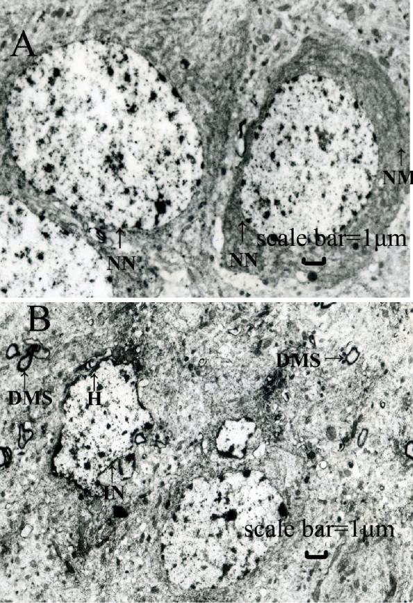
accompanying text reads, "Representative electron micrographs of coronal sections of the rat hippocampus are shown.
(A, C, and E) control hippocampus. (B, D and F) lead-exposed hippocampus in rats at weaning that were treated with 0.2% lead acetate during the gestational and lactational periods as described. Abnormal appearance of neurons including irregular shaped nucleus, swollen mitochondria, often vacuolated with disrupted cristae, a large quantity of heterochromatin collected inside the nucleus, demyelination or shrinkage, and denaturation of the myelin sheath were observed. These findings suggest that hippocampal ultrastructures were injured by lead exposure during the early stage of life. Scale bar = 1 μm. NN: normal nucleus; IN: irregular nucleus; SM: swollen mitochondria; VM: vacuolated mitochondria; NM: normal mitochondria; H: heterochromatin; DMS: denaturation of myelin sheath." (only A and B are used in this modification of the image).
Date
Source
Author
Permission
(Reusing this file)


http://www.jnrbm.com/content/8/1/5 Xu, J; Yan, Hc; Yang,
B; Tong, Ls; Zou, Yx; Tian, Y (Apr 2009). "Effects of lead exposure on hippocampal metabotropic glutamate receptor subtype 3 and 7 in developmental rats" (Free full text). Journal of negative results in biomedicine 8: 5. doi:10.1186/1477-5751-8-5. PMID 19374778. PMC: 2674876. http://www.jnrbm.com/content/8//5.Xu, J; Yan, Hc; Yang, B; Tong, Ls; Zou, Yx; Tian, Y
(modified 9/1/09 by user:delldot)(Reusing this file)
This file is licensed under the Creative Commons Attribution 2.0 Generic license.
- You are free:
- to share – to copy, distribute and transmit the work
- to remix – to adapt the work
- Under the following conditions:
- attribution – You must give appropriate credit, provide a link to the license, and indicate if changes were made. You may do so in any reasonable manner, but not in any way that suggests the licensor endorses you or your use.